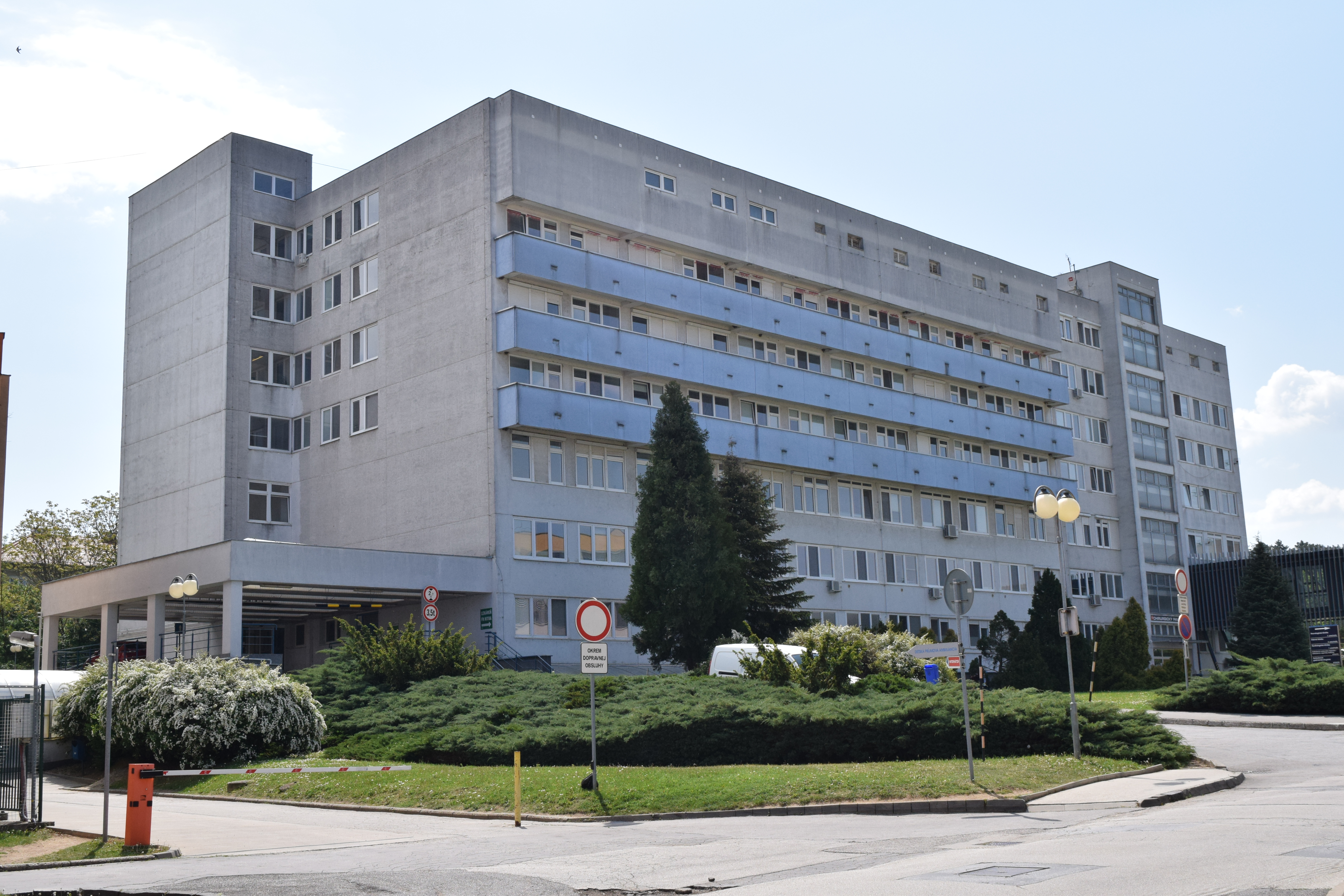
Department of Neurosurgery

The Department of Neurosurgery at Nitra University Hospital was established in 2003. It is currently located on the 5th floor of the Surgical Disciplines Pavilion. The department employs 6 board-certified physicians and 4 physicians in specialization training. The department currently has 22 beds, including 5 monitoring beds, 1 superior-standard bed and 1 semi-superior-standard bed. Approximately 1,000 operations are performed at the Department of Neurosurgery at Nitra University Hospital each year, and more than 7,000 patients are examined and treated on an outpatient basis.

Our Services

The department provides a wide range of neurosurgical procedures for diseases and injuries of the central and peripheral nervous system (in the fields of neuro-oncology, cerebrovascular diseases, neurotraumatology, spine surgery, surgical treatment of hydrocephalus, and pain management including spinal cord stimulation). The range of care provided is further complemented by surgery for certain congenital defects.

  • Neuro-oncological surgery
  • Vascular neurosurgery
  • Spine surgery
  • Neurotraumatology (trauma neurosurgery and neuro-intensive care)
  • Treatment of hydrocephalus
  • Peripheral nerve surgery

Technical Equipment of the Department

Our department is equipped with modern technical equipment, primarily sophisticated instruments used during surgery – an operating microscope, a neuronavigation system, an ultrasonic aspirator, an endoscopic device, and electrophysiological neuromonitoring. The operating room is also equipped with a mobile X-ray machine.

Vascular neurosurgery

Among the most significant and most frequently neurosurgically treated brain diseases are bulges (aneurysms) on cerebral arteries, arteriovenous malformations, and cerebral cavernomas. Neurosurgery also deals with the surgical treatment of bleeding into the brain – intracerebral hemorrhages and certain cerebrovascular accidents.

Cerebral artery aneurysms (bulges) are the most common aneurysm of the arterial system overall. Their prevalence in the healthy population is reported to range from 2–5%. This is a weakening and bulging of the vessel wall of a cerebral artery within the intracranial space. Such a blood-filled bulge of a cerebral artery poses a risk of rupture and subsequent bleeding into the spaces on the surface of the brain (subarachnoid hemorrhage), into the brain parenchyma (intracerebral hemorrhage), or even into the brain ventricles (intraventricular hemorrhage), which manifests as a sudden onset of an extremely severe headache, neck stiffness, photophobia, vomiting, and even impaired consciousness. In the event of bleeding, half of patients die, one quarter survive with neurological impairment, and only one quarter recover and are in good clinical condition.

Several factors contribute to the development of cerebral aneurysms; it is often a combination of a congenital predisposition, and their further development is influenced by atherosclerosis, high blood pressure, and smoking. Their development can also be caused by injuries, infections, or congenital connective tissue disorders.

Treatment of a ruptured or unruptured aneurysm is possible by microsurgical surgery, in which the aneurysm on the given artery is dissected through an opening in the skull using a microscope, and by applying a clip, the aneurysm is excluded from the circulation. The second option is endovascular treatment, in which a catheter is introduced through the femoral artery up to the aneurysm and the aneurysm is filled via the catheter with detachable coils (coiling), or the vessel wall is reinforced (stenting); complex aneurysms can also be treated by inserting flow diverters..

The choice of the most appropriate treatment method for a particular patient is individual, and the decision is the result of an agreement between the neurosurgeon and the interventional radiologist.

Arteriovenous malformationsare a rarer type of vascular anomaly. They are a congenital cluster of inferior vascular structures that may be found incidentally on imaging, or may present with headaches, bleeding, epilepsy, or other neurological symptoms. The choice of the most appropriate treatment method is individual and depends on the size, location, and nature of the blood supply. For some patients, microsurgical removal of the malformation is most appropriate; for others, stereotactic radiosurgery, and sometimes the above treatment methods can be combined with endovascular embolization or occlusion via the vascular system. For some patients, observation alone is most appropriate.

Cerebral cavernomasare low-flow vascular malformations with a mulberry-like appearance, most commonly presenting with epilepsy, headaches, or bleeding. They can occur in any part of the brain. In cavernomas that cause epilepsy or bleeding, surgical treatment is indicated in most cases.

Treatment of chronic pain with spinal cord stimulation

Chronic pain is a problem that can significantly affect quality of life. When basic pain treatment does not work, a modern treatment method – spinal cord stimulation – may be an appropriate option.

What is spinal cord stimulation?
Spinal cord stimulation is a technique that uses gentle electrical impulses to stimulate nerve fibers in the spinal cord area. These impulses help block or relieve pain associated with chronic conditions such as back pain, neuropathic pain, or post-injury pain.

How does it work?
An electrode is implanted in the spinal cord area, and a small battery (pulse generator) is implanted under the skin on the right (or left) side, connected to the electrodes. This device sends electrical impulses that modify how the body perceives pain. The result is pain relief and improved mobility.

Benefits of spinal cord stimulation

  • Pain reduction: Most patients experience significant pain relief.
  • Reduced medication use: Many patients can avoid long-term use of pain medication.
  • Minimally invasive procedure: with a short recovery time.
  • Improved quality of life: Patients report better mobility and reduced discomfort in everyday activities.

Who is spinal cord stimulation suitable for?
Spinal cord stimulation is intended for patients with chronic pain who do not respond to conventional treatment such as medication or surgery. After thorough diagnostics, the physician will decide whether this method is suitable for a particular patient.

Diagnoses for which it is appropriate to consider implantation of a neurostimulator:

FBSS (Failed Back surgery syndrome) patients after repeated lumbar spine surgeries with persistent back and lower limb pain

FNSS (Failen Neck surgery syndrome) patients after repeated cervical spine surgeries with persistent back and upper limb pain

CRPS (complex regional pain syndrome) a chronic painful condition that usually affects a limb after injury or trauma.

Diabetic polyneuropathy of the lower limbs

Ischemic disease of the lower limbs

If you suffer from chronic pain and conventional treatment no longer works, spinal cord stimulation may be an effective solution to help you regain control of your life. Contact our specialists, who will be happy to provide more information and answer your questions.

Spondyloneurosurgery

Spondyloneurosurgery is a branch of neurosurgery focused on the diagnosis and surgical treatment of diseases of the spine and nervous system. It includes surgery of intervertebral discs, degenerative spinal diseases, injuries, tumors, and compression of neural structures. The goal of spondyloneurosurgery is to restore nerve function, eliminate pain, and improve spinal stability so that the patient can safely return to normal activities as soon as possible.

Degenerative changes of the spine

Degenerative changes of the spine are a natural part of aging, but they can also occur at a younger age. There is no single cause; they are often part of complex processes. Gradually, changes occur in the intervertebral discs, vertebrae, and joint structures. Degenerative changes can affect any part of the spine, but the cervical and lumbar spine are most commonly affected, and the thoracic spine rarely.

How do degenerative changes develop?

Over the course of life, intervertebral discs lose fluid, reducing their elasticity. Joints and ligaments may thicken, bony spurs (osteophytes) may form, and the space for the spinal cord and nerve roots begins to narrow. These changes can cause compression of the spinal cord and neural structures, leading to clinical symptoms for which the patient seeks medical attention.

Most common symptoms

Symptoms can vary, but most commonly patients describe:

  • Back pain, which may radiate to the back of the head, head, shoulders, and upper and lower limbs
  • Reduced mobility, muscle weakness, grip disorders, and gait disorders
  • Tingling and sensory disturbances

How are degenerative changes diagnosed?

The basis of diagnosing degenerative changes of the spine is the patient’s medical history and neurological examination. Imaging methods are also an essential part of a properly chosen treatment approach. They can provide important information about the anatomical conditions in a given person; they include:

  • Magnetic resonance imaging (MRI)
  • Computed tomography (CT)
  • X-ray

Treatment options

Treatment is always individual. In most cases, we start with conservative approaches; however, in some cases we proceed immediately to surgical treatment.

Conservative treatment includes

  • lifestyle and movement-habit modification
  • pain and anti-inflammatory medication
  • rehabilitation and physiotherapy
  • injections and infusion therapy

Surgical treatment

Surgery is considered individually based on clinical findings and imaging examinations. In most cases, it is indicated after failure of conservative therapy. At our department, we provide the full range of modern surgical procedures, including:

  • Microscopic surgery for bulging intervertebral discs of the cervical region

The procedure is performed under general anesthesia in the supine position. The surgeon makes an incision in the neck, which is necessary to treat the bulging disc. Then, by dissecting the soft tissues, they reach the pathological disc, which is removed using instruments under visual control of the microscope. After removal of the intervertebral disc, a replacement is implanted. Depending on the extent of the procedure, stabilization of the cervical spine is also performed using a bridging plate fixed into the vertebral bodies.

  • Microscopic surgery for bulging intervertebral discs of the thoracic and lumbar region

The procedure is performed under general anesthesia in the prone position. The surgeon makes a small incision on the spine, which is necessary to treat the bulging disc. Then, by dissecting the soft tissues and bony skeleton, they reach the pathological disc, which is removed using instruments under visual control of the microscope. This approach allows the surgeon to see and manipulate tissue with high precision, reducing the risk of damage to surrounding healthy structures.


  • Decompressive surgery of the cervical, thoracic, and lumbar spine

The procedure is performed under general anesthesia in the prone position. The surgeon makes an incision on the spine, which is necessary to treat compression of neural structures. Then, by dissecting the soft tissues, they reach the bony skeleton. Using instruments, they remove part of the vertebral arch, enlarged joints, and thickened ligaments to ensure the greatest possible freedom for the neural structures.


  • Stabilization surgery via posterior and anterior approaches

PLIF/TLIF – Posterior lumbar interbody fusion/transforaminal lumbar interbody fusion

PLIF and TLIF are very similar surgical techniques, differing in that in TLIF it is not necessary to perform decompression by removing the vertebral arch. These procedures are performed under general anesthesia in the prone position. Through a vertical incision on the back and gradual dissection of soft and bony structures, the surgeon reaches the spine, where they perform decompression necessary to treat compression of neural structures. After removal of the intervertebral disc, a replacement is implanted in its place, and then the procedure continues with insertion of screws into the vertebral bodies and implantation of a stabilization rod that ensures spinal stability.

ALIF

The procedure is performed under general anesthesia in the supine position; the surgical team approaches the spine through the abdominal cavity while continuously protecting the abdominal organs. The pathologically altered disc is removed and a mobile or static replacement is implanted into the resulting space.

  • Biportal endoscopic spine surgery

Biportal endoscopic spine surgery is a modern minimally invasive surgical method performed under general anesthesia in the prone position. In this surgical technique, the surgeon makes 2 skin incisions, using one for the endoscope (camera) and the other for instruments.

Spinal tumors

Spinal tumors are a group of diseases in which abnormal tissue grows in the area of the spine. These tumors can affect the vertebrae, spinal cord, or surrounding tissue. In terms of origin, they can be divided into primary tumors, which arise directly from spinal tissues (primary tumors), or they can spread to the spine from other organs (secondary tumors).

Symptoms of spinal tumors

Symptoms may vary depending on the location and size of the tumor, but most commonly include:

  • persistent back pain that does not improve with rest
  • pain radiating into the limbs
  • weakness, numbness, or tingling in the arms or legs
  • problems with walking or balance
  • urination and bowel movement disorders
  • in advanced cases, loss of mobility

Diagnostics

At our hospital, we use modern diagnostic methods that allow precise determination of the type and location of the tumor:

  • magnetic resonance imaging (MRI)
  • computed tomography (CT)
  • X-ray
  • laboratory tests
  • biopsy

Treatment

Treatment of spinal tumors is a complex process requiring a multidisciplinary approach involving neurosurgeons, oncologists, radiologists, and rehabilitation specialists. The treatment method depends on the type of tumor, its biological behavior, size, location, and the patient’s overall health status.

Surgical treatment
At our department, we provide a wide range of modern surgical procedures aimed at treating spinal tumors. The main goal of surgery is to remove the tumor or part of it, relieve pressure on the spinal cord and nerve roots, and restore spinal stability. This relieves pain, improves nerve function, and prevents further damage.

The basic principles of surgical treatment are:

  • decompression, relieving the spinal cord and nerves from tumor pressure
  • removal of as much of the tumor as possible
  • spinal stabilization

During surgery, we use modern neurosurgical techniques that enable precise, gentle, and safe procedures with an emphasis on minimizing risks for the patient.

Trauma neurosurgery

Head and spine injuries are among the most serious conditions we encounter in medicine. These are life-threatening conditions that require rapid diagnostics, multidisciplinary cooperation, and highly specialized care.

Injuries we treat

At our department, we provide diagnostics and treatment of the complete spectrum of traumatic injuries of the brain and skull and, in cooperation with the Department of Trauma Surgery and Orthopedics, also spinal injuries. Management is based on modern procedures, standardized classification systems, and a multidisciplinary approach.

Craniocerebral injuries – injuries of the skull and brain

Intracranial hemorrhages – we comprehensively treat both acute and chronic bleeding, including:

  • Epidural hematomas (EDH),
  • Acute subdural hematomas (aSDH),
  • Chronic subdural hematomas (chSDH),
  • Intracerebral hemorrhages (ICH).

Skull fractures – linear and depressed skull fractures with a risk of damage to the brain and its coverings, or skull base injury with a risk of CSF leak, damage to cranial nerves, and vascular structures.

Spinal injuries – injuries of the spine, spinal cord, and nerve roots

Spinal injuries – vertebral fractures, traumatic damage to ligaments and intervertebral discs with possible damage to the spinal cord or nerve roots.

Spinal hemorrhages – epidural and subdural hematomas that may lead to spinal cord compression requiring urgent decompression.

Diagnostics of brain and spine injuries

In suspected head or spine trauma, we adhere to the principles of modern management. In addition to clinical examination and neurological assessment, basic imaging examinations are key for rapid and correct diagnosis:

  • CT examination of the brain and spine – first choice in acute trauma.
  • CT angiography – in suspected vascular injury.
  • MRI examination of the brain or spinal cord – for detailed assessment of the extent of damage.

Treatmentof craniocerebral injuries

The goal of treatment of craniocerebral injuries is to prevent secondary brain damage, stabilize vital functions, and, in indicated cases, ensure urgent neurosurgical intervention.

Conservative treatment

In patients without signs of progression of neurological deficit or significant compression of brain tissue, a conservative approach may be chosen, including intensive monitoring of vital functions and neurological status, anti-edema and symptomatic treatment. Repeated imaging examinations to monitor the dynamics of findings are also an important part.

Surgical treatment

Surgical intervention is indicated in worsening neurological status or signs of increased intracranial pressure. The procedures we perform include:

  • Evacuation of intracranial hemorrhages via craniotomy (an artificially created opening in the skull with replacement of the bone after the procedure) or trepanation (a burr hole in the skull)
  • Decompressive craniectomy (removal of part of the skull bone) in progressive increase of intracranial pressure,
  • Cranioplasty (replacement of bone or a titanium implant after decompressive craniectomy),
  • Treatment of depressed skull fractures and injuries of the meninges.

Treatmentof spinal injuries

The goal of treatment of spinal injuries is to prevent secondary damage to the spinal cord and neural structures, restore spinal stability, and maximize the patient’s neurological and functional status. Management takes place in cooperation with the Department of Trauma Surgery and Orthopedics.

Conservative treatment

A conservative approach is indicated in stable spinal injuries without progression of neurological deficit. It includes careful neurological monitoring, immobilization of the spine using braces or fixation aids, and regular imaging follow-ups.

Surgical treatment

Surgical treatment is indicated in unstable spinal injuries, compression of the spinal cord or nerve roots, and in worsening neurological status. We perform:

  • Decompressive procedures (laminectomies, hemilaminectomies) in compression of the spinal cord or neural structures,
  • Stabilization and reconstructive spine surgery using modern fixation systems,
  • Surgical evacuation of spinal hematomas,
  • Treatment of traumatic damage to intervertebral discs.

Postoperative care

Postoperative care is a natural and inseparable part of comprehensive treatment of patients with brain and spine injuries. Its goal is stabilization of vital functions, prevention of secondary damage to the nervous system, and early recognition of postoperative complications.

Patients after neurosurgical procedures are monitored in the monitored beds of our department, or in severe conditions are hospitalized at the Department of Anesthesiology and Intensive Care Medicine (KAIM), where continuous monitoring of vital functions and neurological status is ensured.

Postoperative management includes:

  • Control and treatment of intracranial hypertension and brain edema,
  • Monitoring of neurological status and early diagnosis of complications,
  • Respiratory and hemodynamic support according to the patient’s clinical condition,
  • Prevention of infectious, thromboembolic, and other systemic complications.

After stabilization of the condition, gradual rehabilitation and further treatment follow in cooperation with neurologists, physiotherapists, and other specialized departments, with the aim of maximizing the functional and neurological outcome of treatment.

Peripheral nerve surgery

Diseases of peripheral nerves represent a significant group of neurological disorders that clinically manifest as pain, paresthesias, sensory disturbances, and reduced muscle strength. The most common cause of these difficulties are compressive neuropathies, in which there is long-term or repeated compression of a nerve in anatomically narrow spaces. Untreated compression can lead to irreversible nerve damage and permanent functional deficit.

At our workplace, we focus primarily on the surgical treatment of the most common entrapment syndromes of peripheral nerves of the upper and lower limbs, including:

  • Nervus medianus (median nerve) – carpal tunnel syndrome,
  • Nervus ulnaris (ulnar nerve) – cubital tunnel syndrome,
  • Nervus tibialis (tibial nerve) – tarsal tunnel syndrome,
  • Other compressive neuropathies of peripheral nerves according to individual indication.

Diagnostics of peripheral nerve diseases

The diagnostic process requires a comprehensive assessment of the patient’s clinical condition. The basis is a detailed medical history and neurological examination focused on the distribution of sensory and motor disturbances and provoked compressive signs. Diagnostics are supplemented by ancillary examinations:

  • Electrophysiological examination (e.g., EMG), which objectifies the degree and location of nerve damage,
  • Ultrasound examination of peripheral nerves to assess their morphology and possible compressive changes,
  • MRI examination in suspected secondary cause of compression or in an unclear clinical finding.

Surgical treatment of peripheral nerves

Surgical treatment is indicated in persistent clinical difficulties, progression of neurological deficit, or proven significant nerve compression. The goal of the surgical procedure is nerve decompression, restoration of its physiological conditions, and creation of conditions for regeneration of nerve tissue.

Carpal tunnel syndrome (nervus medianus)

Most commonly manifests as:

  • Tingling, numbness, or burning in the thumb, index finger, and middle finger.
  • Pain in the wrist and hand, often worse at night,
  • A feeling of “clumsiness” of the hand, dropping objects, weakened grip, and impaired fine finger motor skills.

Surgical treatment consists of decompression of the nervus medianus by transection of the ligamentum carpi transversum (transverse carpal ligament). By transecting it, the nerve is released in the carpal tunnel area and there is a gradual reduction of pain and sensory disturbances in the nerve’s innervation area. The operation is most often performed under local anesthesia.

Cubital tunnel syndrome (nervus ulnaris)

Typical symptoms include:

  • Tingling and numbness of the little finger and ring finger,
  • Pain in the elbow area radiating into the forearm or hand,
  • Reduced hand strength and a feeling of stiffness or “disobedience” of the fingers.

The procedure includes decompression (release) of the nervus ulnaris in the elbow area, in indicated cases supplemented by its transposition (relocation). The goal is to eliminate chronic nerve compression and improve motor and sensory functions of the hand. This is an operation performed under general anesthesia.

Tarsal tunnel syndrome (nervus tibialis)

Most commonly manifests as:

  • Pain, burning, or tingling in the sole of the foot or heel area,
  • Numbness of the toes or the entire sole of the foot,
  • Worsening of difficulties with prolonged walking or standing with a feeling of “electric shocks” in the foot.

Surgical treatment consists of decompression of the nervus tibialis and its branches in the tarsal tunnel area. The procedure is aimed at relieving pain, paresthesias, and sensory disturbances in the area of the foot and toes of the lower limb.

Other compressive neuropathies of peripheral nerves

Depending on the affected nerve, the following may occur:

  • Localized pain along the course of the nerve,
  • Sensory disturbances in a specific area of the limb,
  • Reduced muscle strength or decreased coordination of movements,
  • Worsening of difficulties with repeated or long-term movements.

Depending on the location and etiology of compression, we perform targeted decompressive procedures on peripheral nerves of both the upper and lower limbs, including management of postoperative or post-traumatic conditions.

Surgical treatment of peripheral nerves is always indicated individually based on clinical findings and the results of ancillary examinations, with the aim of achieving the maximum possible functional improvement and improving the patient’s quality of life.

What are brain tumors?

Brain tumors are a group of diseases in which there is abnormal growth of cells in the area of the brain, meninges, nerves, or nearby structures. They may arise directly in the brain or spread to the brain from other parts of the body.

Since the brain is enclosed in a rigid skull, even a smaller tumor can cause significant difficulties by compressing surrounding tissue or increasing intracranial pressure. Brain tumors affect children and adults, and their course can be very diverse – from slowly growing and long-term stable to rapidly developing, serious diseases. It is important to emphasize that not every brain tumor is malignant, and modern medicine today offers a wide range of treatment options.

Basic classification of brain tumors

Classification by origin

Primary brain tumors

Primary tumors arise directly from cells of the brain or its coverings. The most common include:

  • Gliomas – tumors arising from the supporting cells of the brain; they may have varying degrees of aggressiveness
  • Meningiomas – tumors arising from the meninges, often slow-growing
  • Pituitary adenomas – tumors of the pituitary gland that may affect hormonal balance
  • Cranial nerve tumors, for example vestibular schwannoma, which may cause hearing and balance disorders

Secondary brain tumors (metastases)

Metastases arise by spread of cancer from other organs (most commonly the lungs, breast, kidneys, skin). They spread to the brain via the bloodstream and are among the most common brain tumors in adult patients.

Classification by biological behavior

  • Non-malignant (benign) tumors
    They grow slowly and do not spread to other parts of the body. Nevertheless, they can cause significant difficulties due to pressure on the brain.
  • Malignant tumors
    They grow faster, often infiltrate surrounding brain tissue, and require complex treatment.

Symptoms of brain tumors

Symptoms of brain tumors depend on:

  • tumor size
  • its location
  • growth rate

The most common manifestations include:

  • headaches, especially new or gradually worsening
  • nausea and vomiting, often unrelated to food
  • epileptic seizures, especially in adults without previous epilepsy
  • weakness of the limbs, gait or coordination disorders
  • speech, vision, or hearing disorders
  • changes in behavior, personality, memory, or concentration
  • disturbances of consciousness or sudden neurological deterioration

Some tumors may be asymptomatic for a long time and only manifest at a more advanced stage.

Diagnostics of brain tumors

Accurate diagnostics are the basis of properly chosen treatment. The aim of examinations is to determine the tumor type, its extent, biological behavior, and relationship to important brain structures.

Imaging examinations

Magnetic resonance imaging (MRI)

MRI is the most important examination in the diagnosis of brain tumors. It provides a detailed image of soft tissues and enables precise assessment of the tumor.

Special techniques are also used:

  • functional MRI (fMRI) – imaging of speech, movement, and other functional areas
  • diffusion tensor imaging (DTI) – visualization of nerve pathways
  • MR spectroscopy – assessment of the chemical composition of tissue
  • perfusion MRI – assessment of tumor blood supply

Computed tomography (CT)

CT examination is fast and accessible; it is used mainly in acute conditions, in suspected bleeding, or in assessment of bony structures of the skull.

PET/CT and functional imaging

PET/CT (positron emission tomography) assesses the metabolic activity of tumor tissue. It helps to:

  • distinguish active tumor from scar tissue or postoperative changes
  • determine tumor aggressiveness
  • monitor treatment effectiveness
  • identify the source of metastases

Biopsy and histological examination

The exact tumor type can only be determined after histological and molecular examination of the tissue. The sample is obtained:

  • during surgery
  • or by stereotactic biopsy, which is minimally invasive

Additional examinations

Diagnostics may also include:

  • neurological examination
  • ophthalmological examination
  • endocrinological examinations
  • neuropsychological examination

Treatment of brain tumors

Treatment is always individual and often combines several treatment approaches.

Neurosurgical treatment –surgery is often the basic step of treatment. The goal is to:

  • complete or partial removal of the tumor
  • reduce pressure on the brain
  • obtain tissue for diagnostics

Modern neurosurgery uses:

  • operating microscopes
  • neuronavigation
  • monitoring of brain functions
  • in some cases awake surgery

Radiotherapy

Radiation is used:

  • after surgery
  • for inoperable tumors
  • for metastases

Chemotherapy and targeted therapy

It is used mainly for malignant tumors and is often tailored to the biological properties of the tumor.

Prognosis and follow-up care

Prognosis depends on:

  • tumor type
  • its aggressiveness
  • extent of surgical removal
  • response to further treatment

After treatment, it is necessary to:

  • regular follow-up examinations
  • long-term monitoring
  • rehabilitation and supportive treatment

Conclusion

Brain tumors are serious diseases, but thanks to modern diagnostic and treatment options, it is now possible to achieve significant improvement in both quality and length of life for patients. Early diagnosis and multidisciplinary cooperation are key to successful treatment.

Contacts

The area code before the extension is 037 / 6545 + extension

Head of Department
Senior physician MUDr. Viktor Šimeg 888
Head nurse Mgr. Mária Nitryová 898
Documentation nurse 989
Ward – nurse 606
Ward – physician 666, 771
Doctors’ room 710
Doctors’ room 386
Meeting Room 682
Neurosurgical Outpatient Clinic 662

Departments

Outpatient Clinics

Our Team

Chief Physician